Gastroenterology image challenge
A 73-year-old woman with a past medical history significant for remote squamous cell carcinoma of the skin presented with a 2-to 3-month history of postprandial dyspepsia and refractory eructation. Laboratory tests were within normal limits. Physical examination was only relevant for mild epigastric pain to deep palpation.
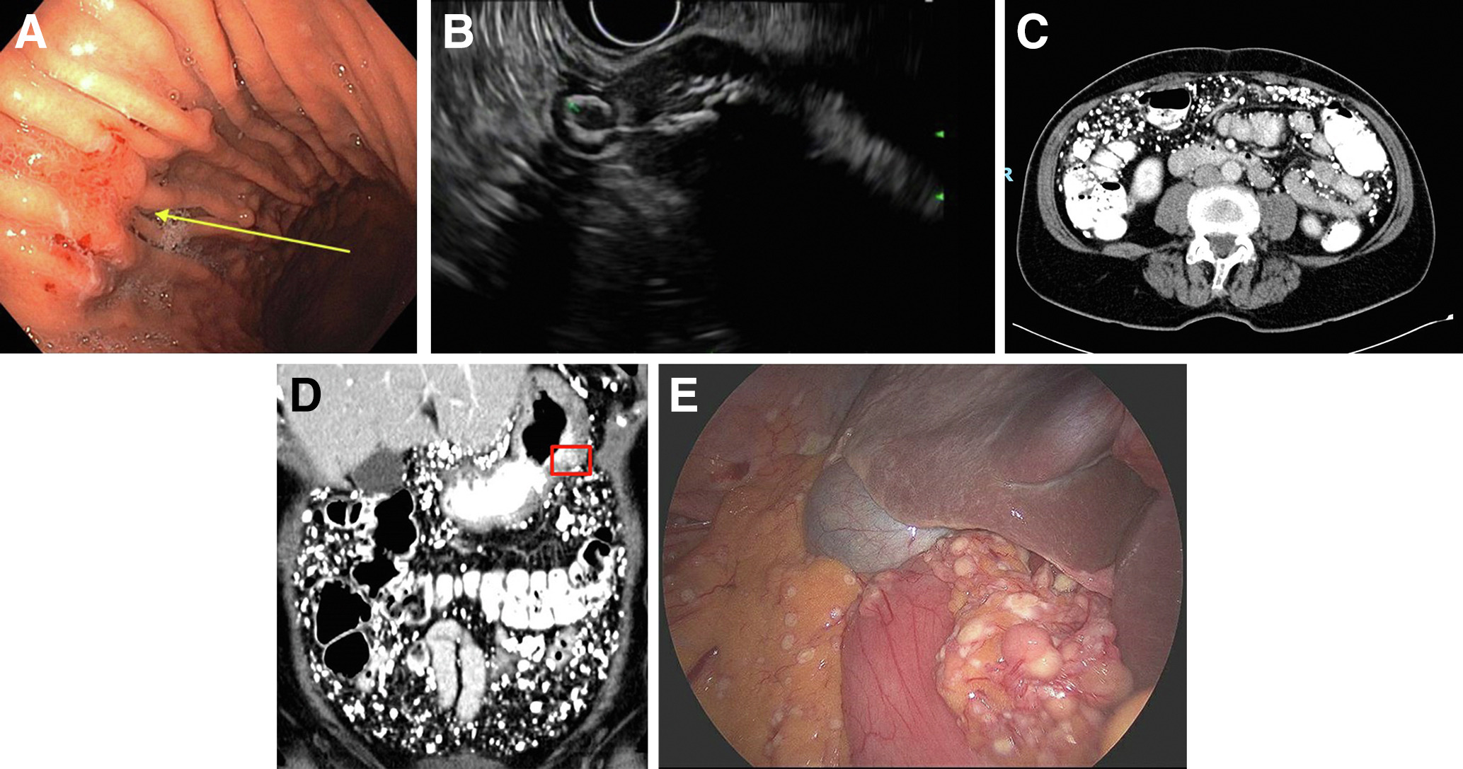
The patient subsequently underwent an EGD that demonstrated localized erythematous and violaceous mucosa along the anterior wall of the gastric body (Figure A). Endoscopic ultrasound evaluation of the abnormality demonstrated a 2.9-cm round calcified subepithelial lesion arising from the muscularis propria (Figure B). A CT scan demonstrated innumerable well-circumscribed rounded calcifications throughout the peritoneum (Figure C, D). Original radiologist read declared highlighted lesion a gastric diverticulum; however, this determination was revised after lesion biopsy results were presented (Figure D), Diagnostic laparoscopy showed similar findings (Figure E).
What is the most likely etiology for the patient’s findings?
To find out the diagnosis, read the full case in Gastroenterology.


