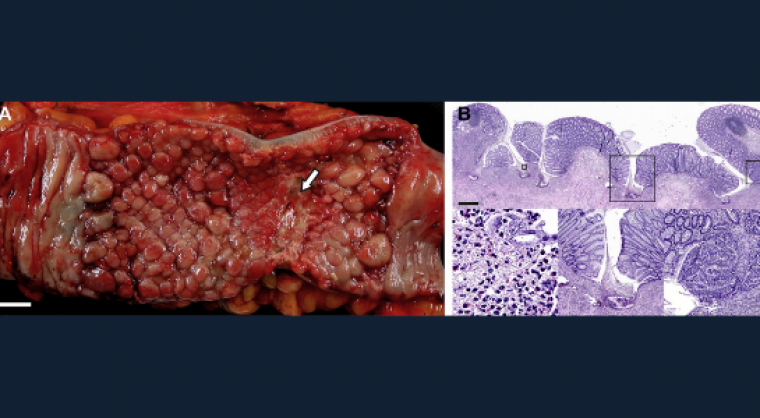
Gastroenterology image challenge: A 41-year-old woman underwent sigmoid colectomy. As occurs commonly, the specimen was received in the surgical pathology laboratory with no information other than the patient’s name, medical record number and “sigmoid.” The surgeon could not be contacted. On opening the colon, a well-defined region with innumerable small polyps was identified (figure A). Within this region was an ulcer and associated stricture. Proximal and distal mucosal margins were uninvolved and had normal folds.
Histologic sections were taken and, at low power, the polyps were noted to be small and regularly spaced (figure B). The small, regularly spaced polyps were separated by well-defined zones of ulceration that began within the stalk and extended into the superficial submucosa (figure B). The inflammatory infiltrate within the ulcers was composed of eosinophils (figure B) with admixed neutrophils. Most polyps were composed of normal-appearing mucosa, but a few had a different appearance (figure B).
What is the diagnosis and what additional information should the pathologist provide?
See the full case in Gastroenterology.


