Gastroenterology image challenge:
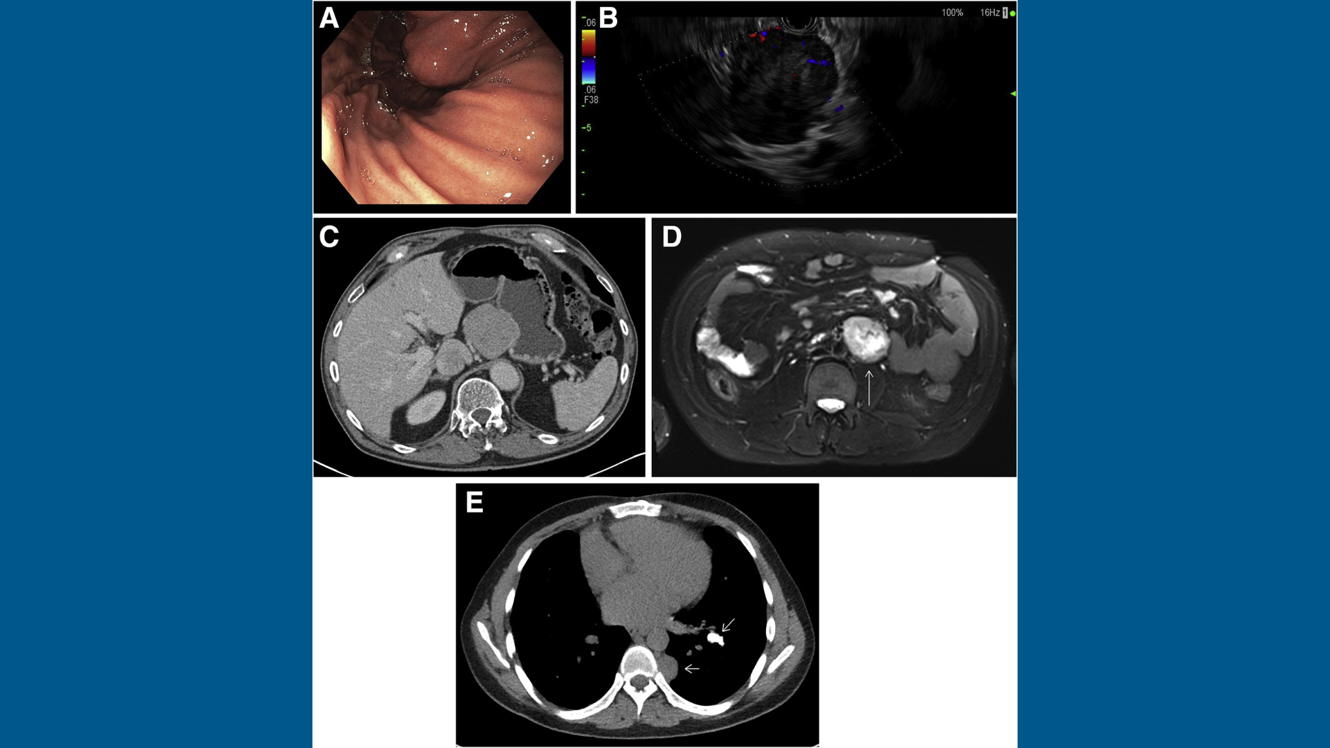
A 26 year-old man complained of vague abdominal pain along with episodes of intermittent melena. He was otherwise healthy with no relevant past medical history and was taking no medications. The patient underwent upper endoscopy with ultrasound which detected a mass along the lesser curvature of the stomach (Figure A, B), which was biopsied. A computed tomography scan of the abdomen was performed after endoscopy (Figure C), which detected a para-aortic mass that was further studied with magnetic resonance imaging (Figure D).
At that point, there was concern for a possible malignant process, so a computed tomography scan of the chest was done that detected calcified lung nodules and a paraspinal mass (Figure E).
What diagnosis explains the constellation of findings in this young patient?
To find out the diagnosis, read the full case in Gastroenterology.


