


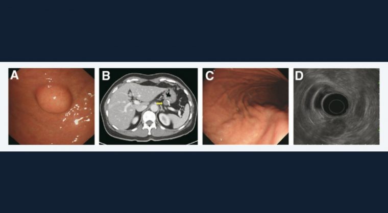
Gastroenterology image challenge: A 58-year-old woman presented with a subepithelial tumor, approximately 2 cm in size, incidentally detected in the posterior wall of upper body of the stomach during a screening endoscopy (Figure A). The sample from an endoscopic ultrasound-guided fine-needle biopsy revealed spindle-shaped cells arranged in a fascicular pattern. The spindle cells were immunopositive for c-Kit, cluster of differentiation 34, DOG-1, and smooth muscle actin.
The patient was diagnosed with a gastrointestinal stromal tumor (GIST) and underwent laparoscopic wedge resection. The tumor was completely resected with clear margins. The maximal tumor size was 2.3 cm, and the mitotic count was 2 per 50 high-power fields; therefore, it was diagnosed as a GIST with a low risk of malignant potential.
Follow-up abdominal computed tomography after 2 years showed a new low-density mass in the previous resection site (Figure B, arrow). Endoscopy showed no definite lesion at the resection site (Figure C), and endoscopic ultrasound revealed a 1.5-cm heterogeneous hypoechoic lesion originating from the muscularis propria (Figure D). Laparoscopic wedge resection was performed again for a definitive diagnosis and treatment.
What is the most likely diagnosis? How should the patient be managed?
To find out the diagnosis, read the full case in Gastroenterology.