


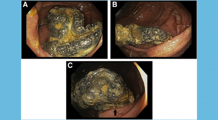

Gastroenterology image challenge: A 61-year-old man with known diverticulosis and a recurrent ventral hernia requiring multiple open surgical repairs reported a single episode of self-limited hematochezia. He denied abdominal pain, nausea, vomiting, altered bowel habits, rectal pain, fecal urgency, weight loss, fever, or chills. He had a remote smoking history with excessive alcohol consumption. The physical examination was entirely unremarkable. Laboratory investigations were also unrevealing, including normal hemoglobin (14.1 g/dL) and mean corpuscular volume (85.9 fL). Colonoscopy, performed to identify the source of hematochezia, revealed a large (4- to 6-cm long), firm, blackish foreign body in the cecum (Figure A). On closer inspection and manipulation of the foreign body with a Roth net and snare, mucosal ulceration was noted and it became apparent the foreign body was firmly attached to the cecal wall; it was not removed (Figure B, C). The remainder of the endoscopic examination revealed severe nonbleeding diverticulosis in the descending and sigmoid colon and small nonbleeding internal hemorrhoids.
What is the most likely diagnosis and what are the next steps in management?