News
Featured Articles
The Shark Tank winner is Arithmedics!
Have you downloaded the I-SEE mobile app yet?
Top basic science sessions to attend at DDW
Calling all clinicians: mark your calendar for these DDW sessions
New opportunity for patients to share about iron deficiency anemia
New HHS guidance on informed consent impacts GIs
Check out our new Crohn’s disease clinician toolkit
Trainees: Take advantage of DDW® sessions and meetups
Let’s mingle at DDW®

AGA Pocket Guides
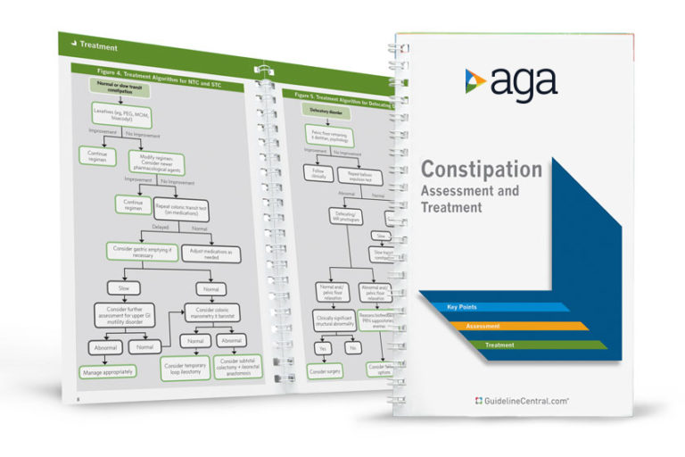
Official AGA Institute quick-reference tools provide healthcare providers and students with instant access to current guidelines and clinical care pathways in a clear, concise format. AGA Institute pocket guides are available in print and digital form.
Member Non-Member
AGA clinical guidance
Find the latest evidence-based recommendations for treating your patients.


