News
Featured Articles
Discover inspiring research at DDW® 2024
3 weeks left to register for DDW®
Join AGA for international education opportunities at DDW® 2024
How to navigate mentorship
Review your reported provider data in Open Payments
The Shark Tank winner is Arithmedics!
Have you downloaded the I-SEE mobile app yet?
Top basic science sessions to attend at DDW
Calling all clinicians: mark your calendar for these DDW sessions

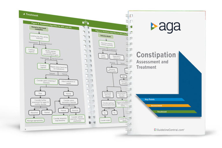
AGA Pocket Guides
Official AGA Institute quick-reference tools provide healthcare providers and students with instant access to current guidelines and clinical care pathways in a clear, concise format. AGA Institute pocket guides are available in print and digital form.
Member Non-Member
AGA clinical guidance
Find the latest evidence-based recommendations for treating your patients.


